Clinical Pathology

Clinical Pathology Service provides standard and customized research grade and clinical grade histology and pathology services including routine and special histological stains as well as immunofluorescence and immunohistochemistry. Clinical specimens are protected by HIPPA privacy rule. Deidentified patient data are deposited and transmitted via a protected secure server.

Background

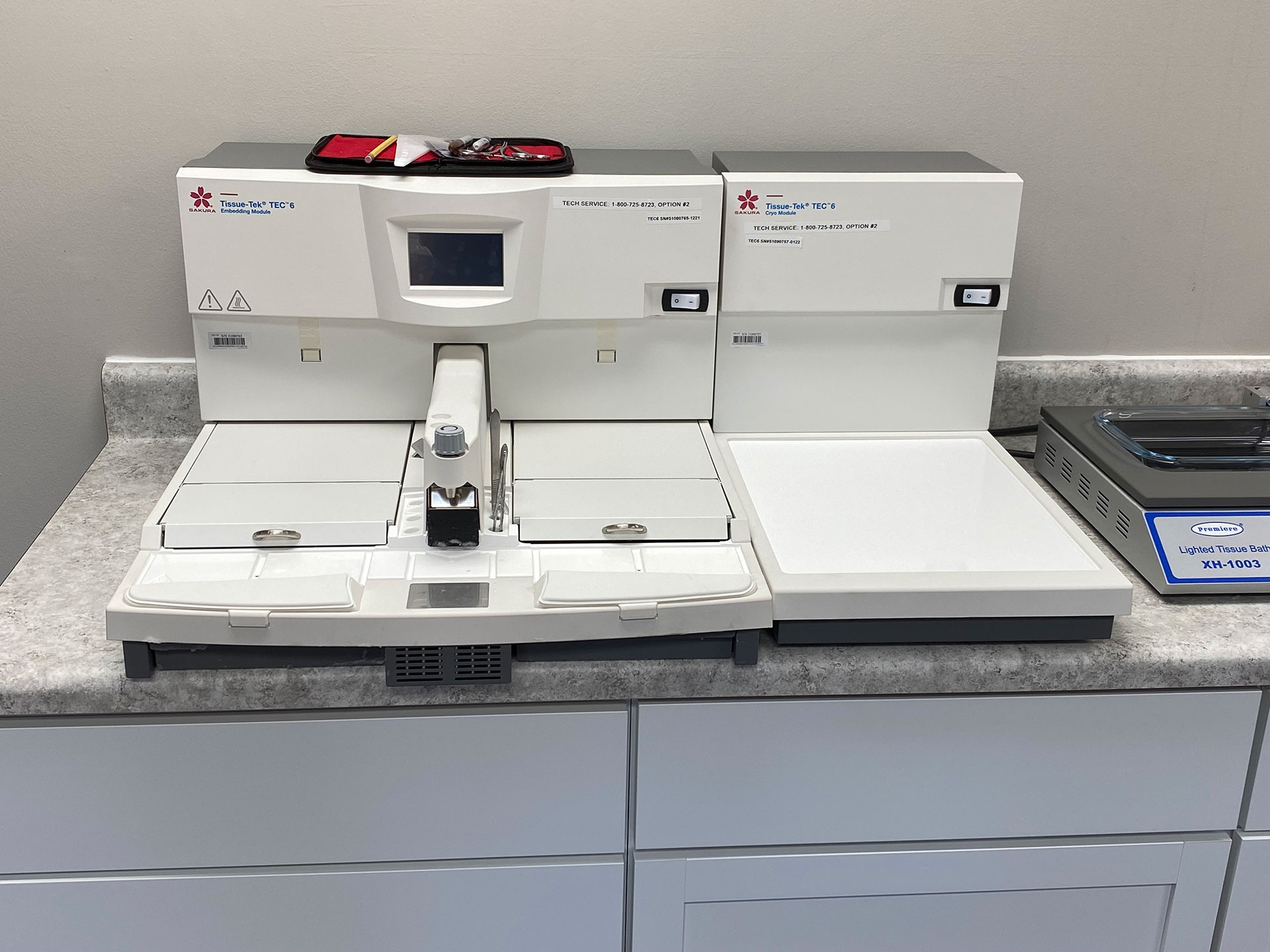
Our clinical pathology service provides standard and customized research-specific histology and pathology services including routine formalin fixation, and paraffin embedding and sectioning, and cryosectioning of frozen sections.

Clinical pathology offers routine and special histological stains, immunohistochemistry and immunofluorescence, and in situ hybridization against mRNA or microRNA, as well as digitized imaging data collection using Nikon Ni-E motorized microscope equipped with CCD fluorescence camera and bright-field color camera.

We have worked with a wide variety of tissues and staining techniques from mice, rats, non-human primates, pigs and humans and can develop protocols for new antibodies.

bicell-blue-clinical-pathology-equipment04

Mice and rats

Non-human primates

Pigs

Humans

Services

Tissue processing and embedding

Sectioning and staining of paraffin-embedded and cryostat sections

Routine and special histological stains
See “Sample Submissions” for the list of stains

Immunohistochemistry (DAB or other labels including fluorescent labels)
See “Sample Submissions” to select an antibody from BiCell Scientific catalog

Immunofluorescence (Alexa Fluor 488 or other colors)
See “Sample Submissions” to select an antibody from BiCell Scientific catalog

In situ hybridization (DIG probes against mRNA or microRNA)
See Sample Submissions to select a mRNA or microRNA probe

Digital imaging using Nikon Ni-E motorized microscope

bicell-blue-clinical-pathology-equipment

Turnaround time

  • Small orders of 10 samples or less – 5 days
  • Large orders of greater than 10 samples -2 weeks

Deliverables

Each sample will have 2 consecutive sections on one slide with specified stain or label. Slides will be examined with light microscopy or epifluorescence microscopy. Slides with meaningful signals will be shipped to customers.

Imaged micrographs will be sent to customers via BiCell Blue Cloud.

Ready to get started?

Order from a range of options today!

Scroll to Top